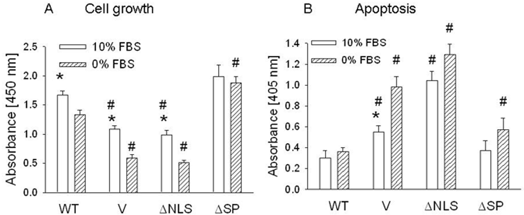
Figure 2. Cell growth (A) and apoptosis (B) of LoVo cells overexpressing wild-type (WT) PTHrP or PTHrP deleted over the nuclear localization signal (ΔNLS) or signal peptide (ΔSP).

V = empty vector control. Cells were plated in 10% FBS. Cell growth and apoptosis under serum replete conditions were measured 96 h and 72 h after plating, respectively. For experiments under serum-depleted conditions, cells were transferred to 0% FBS 24 h after plating. Cell growth and apoptosis were measured after a further 72 h and 48 h, respectively. Each bar is the mean ± SEM of three independent experiments for each of three independent clones. * = Significantly different from the corresponding 0% FBS value (P < 0.001); # = significantly different from the corresponding WT value (P < 0.001).
