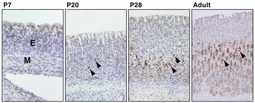
Figure 2.
Ontogeny of apelin immunoreactivity in the oxyntic mucosa of the developing rat stomach. Apelin-containing cells (brown color) were not detected at P2 (not shown), P7 and P13 (not shown) and were observed initially in the rat stomach at 20 days of age (P20) by IHC. Apelin-containing cells are localized primarily in the middle and basal region of gastric glands (arrow heads). The density of apelin-containing cells increased progressively with age. Omission of apelin antibody or use of control rabbit serum in place of the rabbit apelin antibody failed to detect apelin-positive cells (not shown). Photos were captured with a 10× objective, M = muscle layer of stomach; E = gastric mucosal epithelium; P7 = postnatal day 7.

