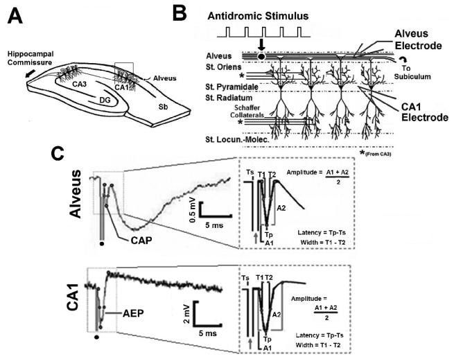
Fig. 1. Field potentials evoked by hippocampal fiber tract stimulation in-vitro.
A. In-vitro experimental schematic. Sb – Subiculum, DG- Dentate Gyrus. B. Extracellular field potentials were recorded simultaneously in the CA1 stratum pyramidale (CA1 electrode) and alveus (Alvear Electrode). C. Simultaneously recorded compound action potential (CAP) and antidromic evoked potential (AEP) in-vitro in response to antidromic stimulation of the alveus. Dot denotes stimulus artifact. Inset: AEP and CAP amplitude, width, and latency measurements.

