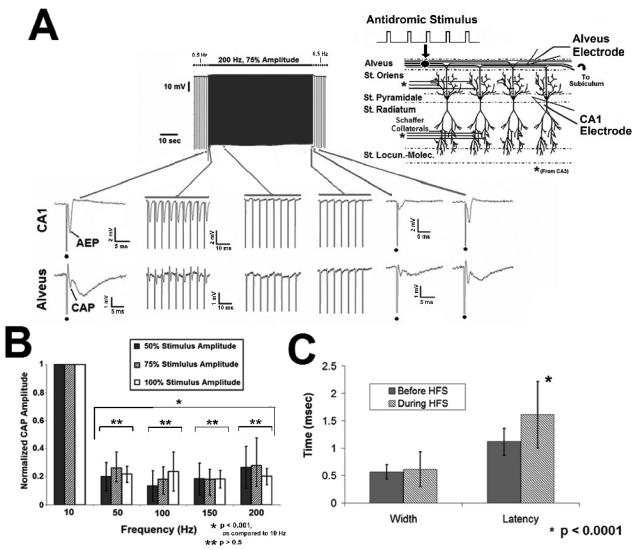
Fig. 3. Evoked axonal responses fail to follow HFS pulse trains in-vitro.
A. Evoked responses (CAP) failed to follow prolonged pulse train HFS. The frequency of the evoked stimulus given before and after HFS was 0.5 Hz. The example illustrates the effect of a 200 Hz pulse train. HFS generates initial excitation that decreases in amplitude over the stimulus duration, until the evoked response is gone. Dot denotes stimulus artifact during 0.5 Hz control stimulation. B. Failure of HFS evoked excitation is frequency dependent (p<0.001, ANOVA), with responses unable to faithfully follow pulse train HFS above 50 Hz. Effects on somatic activity (AEP) were similar (not shown). C. CAP width and latency increased prior to complete failure (mean +/− SD; p<0.016, p<0.0001, respectively, student's paired t test), AEP width and latency were unchanged (p<0.2, student's paired t test; not shown).

