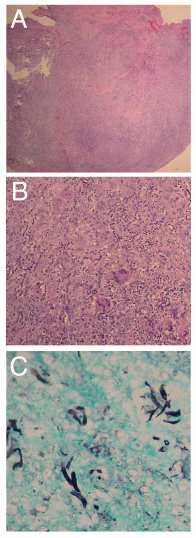
Figure 2. Histology.
A. Low power image of the tissue biopsy showing a nodular fragment of fibroadipose tissue with chronic lymphohistiocytic infiltrate, necrosis, and fibrosis (hematoxylin and eosin, 20X). B. Higher power view of the same specimen demonstrating a dense granulomatous reaction with multiple foreign body-type giant cells (hematoxylin and eosin, 100x). C. GMS stain shows fungal hyphae with 45° branching (200x).

