
Figure 1. PTEN expression in VHL null cells correlates with increased expression of the HIF-2α target, MT1-MMP.
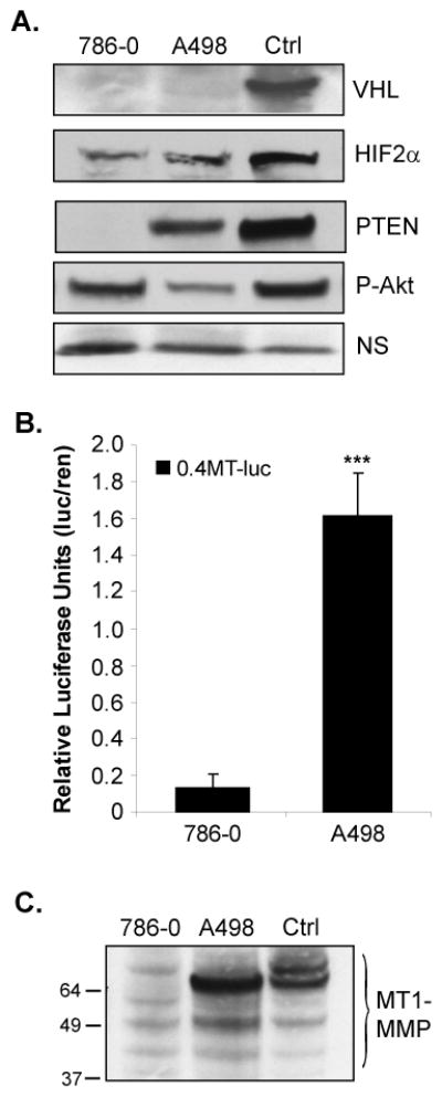
A. Whole cell lysates of the human RCC cell lines, 786-0 and A498, were analyzed by western blotting for expression of VHL, HIF-2α, PTEN, and P-Akt (ser 473). As a positive control (Ctrl) for immunoblotting purposes only, the 786-0 derivative line (WT8) was transfected to express VHL, HIF-2α, or PTEN. The WT8 cells have endogenously high levels of P-Akt, due to lack of PTEN expression. A non-specific (NS) band was used as a loading control. B. 786-0 and A498 cells were transiently co-transfected with 0.4kb of the human MT1-MMP promoter upstream of luciferase (0.4MT-luc) and pCMV-renilla. Cells were harvested and analyzed by the Dual Luciferase Assay®. Results represent three independent experiments of triplicates samples and are presented as relative luciferase units (luciferase normalized to renilla). C. MT1-MMP protein expression in whole cell lysates of the 786-0 and A498 lines as analyzed by western blotting. As a control, WT8 cells were transfected with the MTpc3SE construct to express MT1-MMP as previously described (12). Note that MT1-MMP protein appears as several cleavage products, which is not unusual for this protein (70). Results represent the average of three independent experiments of triplicates samples. Protein expression studies were repeated at least three times. Columns, mean; bars, SD; p<0.0001.