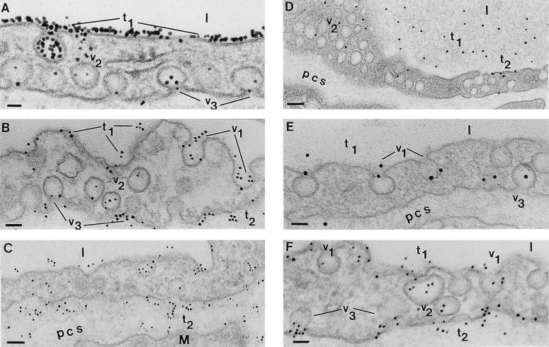
Figure 2.
Postembedding immunodetection of Om-DNP in perfused and bolus-administered specimens. (A) Capillary profile in a specimen perfused for 5 min with Om-DNP (at 5 mg/ml). The luminal endothelial surface is extensively labeled by the tracer (t1). Inside the endothelium only a few caveolae (v2) are labeled, but even at this early time, caveolae (v3) discharging their content could be seen. l, lumen. (Bar = 35 nm.) (B) After 15 min Om-DNP molecules are associated with the luminal plasmalemma (t1), with caveolae opened to the lumen (v1), with caveolae apparently free in the cytosol (v2), and with caveolae that are discharging their content into the pericapillary spaces (v3). Note also the presence of apparently free tracer molecules (t2) in the perivascular spaces. (Bar = 60 nm.) (C) As time of perfusion progresses (30 min), the tracer is found all over the endothelial profile with the same distribution as in B, but the label density (t2) over the perivascular spaces is much more extensive. M, muscle; pcs, pericapillary space(s). (Bar = 84 nm.) (D) After 5 min of an i.v. injection of 100 μl from a concentrated Om-DNP solution (of 50 mg/ml), the tracer is present in the lumina of the vessels (t1), associated with the plasmalemma proper (t2), with the caveolae opened to the luminal and abluminal (v3) side of the endothelium, and with caveolae apparently free in the cytosol (v2). (Bar = 100 nm.) (E) At 15 min from bolus administration of Om-DNP the majority of the tracer is still found in the lumen (t1). The tracer molecules label more extensively caveolae opened to the lumen (v1) as well as caveolae that are discharging their content (v3) in the perivascular spaces. (Bar = 52 nm.) (F) Thirty minutes after i.v. administration, the tracer is still found in the lumina (t1) but many caveolae opened to the lumen (v1) or scattered inside the cells (v2) are labeled, as are the caveolae (v3) discharging on the abluminal front of the cell; some of the latter contain multiple tracer molecules. Note the extensive labeling (t2) of the perivascular spaces at this time, resembling that found in perfused specimens; yet its density remains much lower than that encountered in perfused specimens (compare with C). (Bar = 53 nm.)

