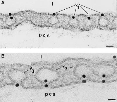
Figure 3.
Binding pattern of Om-Au. (A) When Om-Au (≈22 nm diameter) was perfused as in Fig. 2 A–C, the necks of many caveolae opened to the lumina (v1) were plugged by the tracer. (Bar = 49 nm.) (B) By 15 min of perfusion, the gold coated particles are seen plugging the necks of the caveolae (v3) opened on the abluminal front of the cells. Not so often, the necks of some caveolae (v3) opened on the tissular side of endothelial cells are plugged with two gold particles. (Bar = 28 nm.) l, lumen; pcs, pericapillary space(s); the same abbreviations are used for Fig. 4.

