
Figure 1.
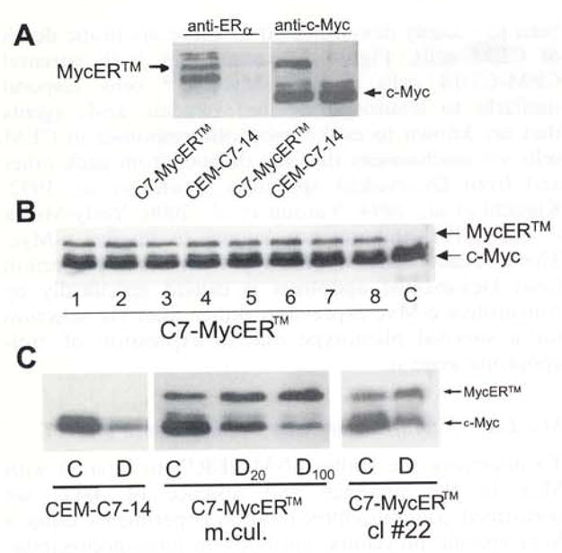
Expression of MycER™ in CEM-C7-14 cells. CEM-C7-14 cells were stably transfected with a plasmid pBpuroMycER™, selected for puromycin resistance and cloned in 0.5% soft agar. Individual colonies were isolated and amplified. Fifty microgram total cellular protein extracted from uncloned mass culture, individual clones or the parental untransfected CEM-C7-14 cells (C7) was resolved by SDS–PAGE, electroblotted, and the membranes were analysed by Western blotting. (a) Extracts of CEM-C7-14 cells and uncloned mass cultures of C7-MycER™ cells probed with an anti ERα antibody recognizing an epitope corresponding to the hinge region of ERα, or an anti-c-Myc monoclonal antibody Mycl-9E10.2) (b) MycER™ expression (antibody Mycl-9E10.2) in eight representative clones. (c) The parental CEM-C7-14 cells, uncloned C7-MycER™ (designated: m.cul.) and a representative clone (cl #22) of C7-MycER™ cells were treated with 20 nM Dex (D20), 100 nM Dex (D or D100) or ethanol vehicle (C) for 24 h. Whole cell extracts corresponding to 50 μg protein were subjected to SDS–PAGE and Western blotting using the anti-c-Myc antibody Mycl-9El0.2