
Figure 7.
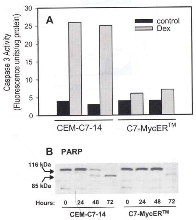
Caspase activation in Dex-evoked apoptosis. (a) Caspase 3 activity was measured using the substrate Z-DEVD-AFC in extracts of CEM-C7-14 and C7-MycER™ cells treated for 48 h with ethanol (solid bars) or 100 nM Dex (gray bars). Data are from a representative experiment, which consisted of two independent treatments; each is presented as a separate bar. Each bar represents an average of duplicate measurements. (b) CEM-C7-14 and C7-MycER™ cells were treated with 100 nM Dex for the indicated length of time, harvested, and whole cell extracts were prepared. Aliquots corresponding to 50 μg protein were resolved by SDS–PAGE, electroblotted, and the membrane was probed using an antibody (sc-7150, Santa Cruz Biotech.) that reacts with the uncleaved as well as cleaved PARP proteins