
Figure 1.
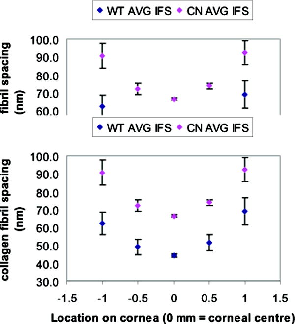
Graphical representation of average collagen interfibrillar (Bragg) spacing (y-axis) from X-ray scattering patterns obtained at 5 sites at 0.5 mm intervals (x axis) along a vertical meridian across the cornea in 5 wild type and 8 Klf4CN mice. In both wild type and Klf4CN stroma, the interfibrillar spacing becomes increasingly greater on moving further towards the periphery from the corneal centre (represented by 0mm). Fibril spacing in the Klf4CN mouse cornea exceeds that in wild type across the meridian.