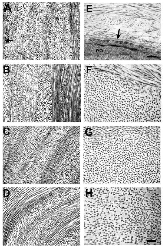
Figure 2.

Electron micrographs of corneal lamellae in wild type (A-D) and Klf4CN corneal stroma (E-H). Subepithelial stroma appears disrupted in mutant cornea (E); arrows indicate basal lamina below epithelium; anterior stroma (B and F); mid stroma (C and G); posterior stroma (D and H). Bar = 480nm (A, E), 300nm (B-D, F-H).
