
Fig. 6.
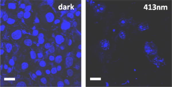
Confocal fluorescence microscope images of the PANC-1 cancer cells that were treated with a suspension of the nanoimpeller-functionalized particles loaded with the camptothecin (CPT), kept in the dark (left) and 5 min illuminated at 413 nm of a light (right). Photoactivated impellers released the CPT from the particles inducing cell apoptosis (right) while unexcited machines did not cause the CPT release and cells were intact (left). Scale bar: 30 μm.