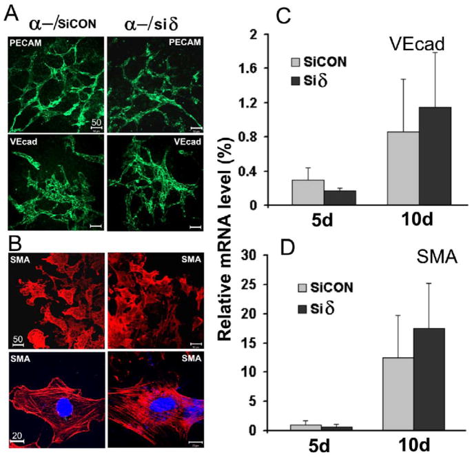
Fig. 4. Differentiation of ESCs to ECs and SMCs.
EB-monolayers derived from p38α−/−/siCON ESCs (α-/siCON), and p38α−/−siδ ESCs (α-/siδ) were immunostained with; A, antibodies against EC markers PECAM-1 (PECAM) or VE-cadherin (VEcad) followed by FITC-conjugated secondary antibodies (green); B, antibodies against SMC marker SMA followed by rhodamine-conjugated secondary antibodies (red). The cells were examined and photographed with a confocal microscope (scale bar unit =μm). C & D, quantitative determination of VEcad and SMA expression by RT-qPCR. The mRNA of each gene was determined from 5 and 10 day EBs and normalized to β-actin mRNA. The expression level of β-actin was set as 100 %. Results are means ± SEM of three independent experiments.

