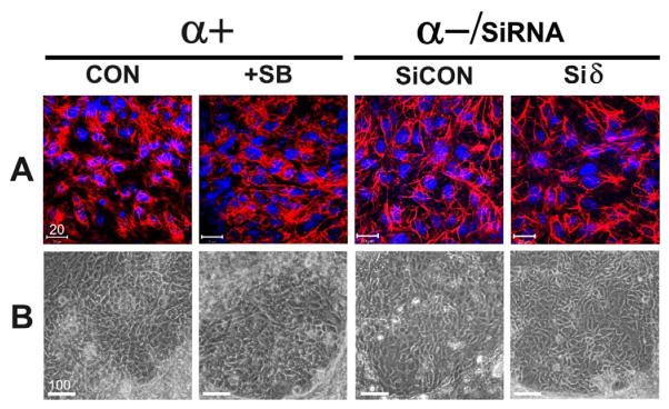
Fig. 5. Differentiation of ESCs to keratinocytes.
EB-derived monolayers were differentiated from ESCs of the following genotypes: p38α+/+ESCs without treatment (p38α+, CON); p38α+/+ ESCs in the presence of 5 μM SB (p38α+, SB); p38α−/−/siCON ESCs (α-/SiCON), and p38α−/−siδ ESCs (α-/siδ). A, keratinocytes identified by immunocytochemistry. Differentiated cells were immunostained with an antibody against cytokeratins (C-2562, Sigma) followed by rhodamine-conjugated secondary antibodies and photographed under a confocal microscope. B, the cytokeratin positive cells identified in A were examined and photographed under a phase contrast microscope. These cells were detected in “patch-like” areas and have typical morphology of keratinocytes (scale bar unit = μm).

