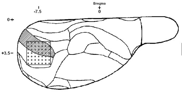
Figure 1.
Outline of electrode placement as viewed from the dorsal surface of the right hemisphere of the rat brain. Each dot represents an electrode penetrating at approximately a right angle to the brain surface. Coordinates indicate the location of the center of the electrode array relative to Bregma. Shaded area is the monocular region of the primary visual cortex. The region immediately lateral is the binocular region of the primary visual cortex.

