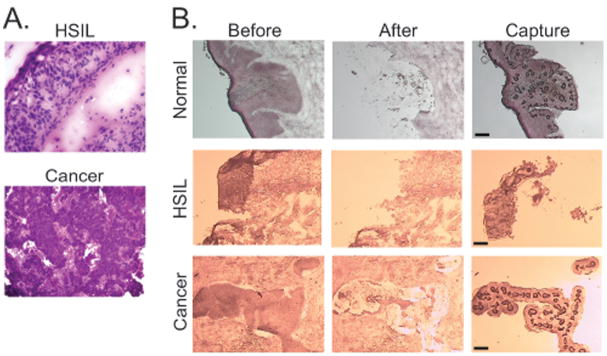
FIG. 2. Representative histological sections.
A. Histopathology of HSIL and cancer specimens. Prior to performing LCM, pilot sections were stained with hematoxylin and eosin to screen for lesional tissue (HSIL, 200X; cancer, 100X). B. Typical LCM results. Frozen sections of normal cervical epithelium, high-grade squamous intra-epithelial lesion (HSIL) and cervical squamous carcinoma (cancer) were stained with Nuclear Fast Red. Microscopic images before LCM and after LCM are shown along with the annealed tissue captured on the cap, Dark circles in the captured tissue represent sites of direct laser energy deposition, where the cap polymer annealed to the underlying tissue. Scale bar, 30 μm.

