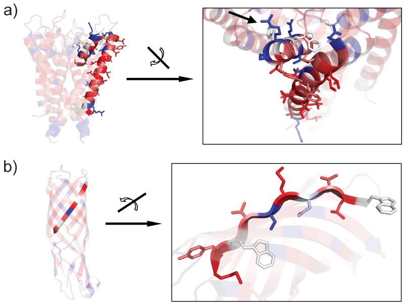
Figure (7).
Close-ups of Figure (6) a) and d). The figure demonstates the ability of the UHS to correctly identify the structural context of the amino acids within a functional protein. Figure (7a) displays the prediction for the pore helix of the KcsA potassium channel (short helix on the top). Figure (7b) demonstrates that the UHS is clearly able to distinguish the different hydrophobicities of the side-chains in the β-barrel. More details are given in the Results and Discussion section.

