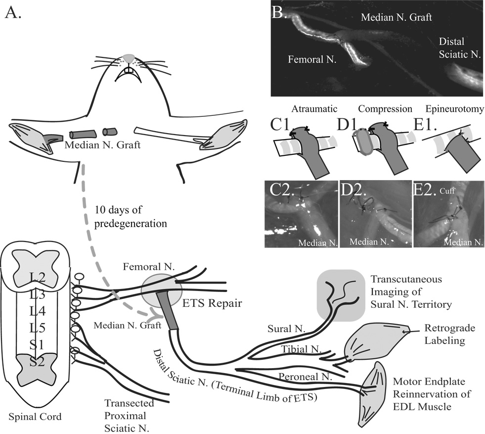
Figure 2. Schematic of experimental design.
A. The median nerve was proximally transected 10 days prior to ETS repair to eliminate YFP- or GFP-labeled axons. The 5 mm median nerve graft was coapted ETS to the common femoral nerve proximally, and end-to-end into the distal stump of the transected sciatic nerve distally. Motor endplate reinnervation of extensor digitorum longus (EDL), retrograde labeling of the gastrocnemius muscle to the L2–L4 spinal cord, and cutaneous reinnervation of the sural nerve territory were evaluated. B. Live image at t=0, with a fluorescence naïve median nerve graft spanning the femoral nerve proximally, and the sciatic nerve distally in a mThy1-GFP(S) mouse. The labeled axons in the transected sciatic nerve also degenerated to facilitate axon tracking. C1. Schematic of “atraumatic” repair. C2. Brightfield image. D1. Schematic of “compression” repair. D2. Brightfield image. E1. Schematic of “epineurotomy”. E2. Brightfield image.

