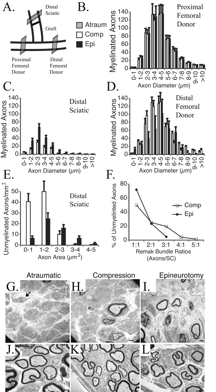
Figure 6. Morphometric and stereologic assessment of donor and recipient nerve cross sections.
A. Donor nerves were sectioned 5 mm proximal and distal to the ETS repair and in the reconstructed limb of the transected sciatic nerve 5 mm distal to the median nerve graft. B. The proximal femoral donor nerve demonstrated a consistent distribution of myelinated axons counts based on fiber widths. C. The reconstructed sciatic nerve demonstrated the most axons with epineurotomy repair, and none with atraumatic repair. D. The distal femoral nerve demonstrated progressively fewer myelinated axons from atraumatic to compressive to epineurotomy ETS repair. E. Compressive repair resulted in the highest total number of unmyelinated axons, particularly with areas ≤2 µm2. No unmyelinated axons were noted with the atraumatic repair. F. A higher percentage of unmyelinated axons were associated with larger Remak bundles with compressive than epineurotomy repair. G. EM of sciatic nerve reconstructed with atraumatic; H. compressive; I. epineurotomy repair. Arrow points to scant unmyelinated axons that lack a double basement membrane or SCs. J. Distal femoral donor nerve reconstructed with atraumatic; K. compressive; L. epineurotomy ETS repair. In compressive repair (K.), note the particularly large Remak bundles with up to 47 unmyelinated axons per SC distal to the compressive ETS repair. Scale bars 1 µm.

