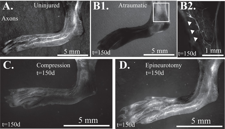
Figure 8. Transcutaneous imaging of the sural nerve.
A. Uninjured control shows sural nerve appearing inferior to the hamstring and innervating the lateral lower leg, ankle, and skin over the dorsal fifth metatarsal of the foot. B1. With atraumatic ETS repair, no sciatic nerve branches are visualized at 150 days at low magnification. B2. At higher resolution, a few YFP-labeled cutaneous axons (white arrowheads) were noted, in one mouse in the sural nerve territory. C. Scant reinnervation of a few peroneal branches on the dorsum of the foot, but no clear sural nerve branches with compressive ETS repair. D. Restoration of sural nerve (and visible peroneal nerve) with epineurotomy repair by 150 days.

