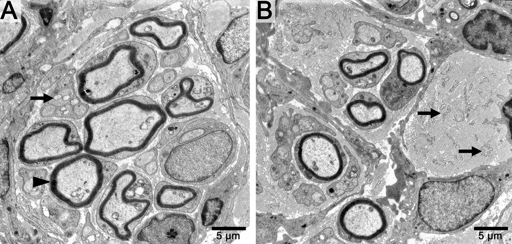
Figure 3.
Electron micrographs of distal tibial nerve sections in cold motor and cold sensory nerve groups. A. Note the multiple myelinated (black arrow head) and unmyelinated fibers (black arrow) in the motor group. B. The sensory nerve groups show fewer fibers and multiple empty bands of Bungner which contain Schwann cell lamina tubes (black arrows). Scale bar in bottom right hand corner.

