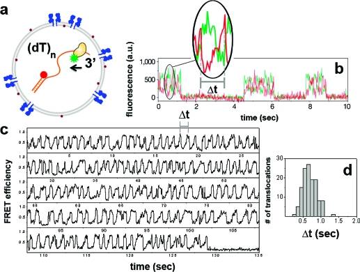
Figure 4.
(a) Rep helicase labeled with donor, and DNA labeled with acceptor at the junction depicted as coencapsulated within a porous SUV. Partial duplex DNA is an 18bp double strand carrying a (dT)80 tail. (b) Rep shuttling data obtained from surface-tethered DNA (modified from ref (32)) with a same length tail. Inset shows a single translocation event where the gradual increase in the acceptor signal (with accompanying decrease in the donor signal) reflects translocation of Rep toward the junction, whereas the abrupt drop marks the snapping back to the 3′ end. The translocation time (Δt) is defined as the period between consecutive snapping events. Individual cycles of shuttling separated in time are due to different Rep molecules from solution binding on the same surface-attached DNA and exhibiting limited repetitive translocations until dissociation. (c) FRET trace of Rep shuttling on a (dT)80 DNA within 100 nm diameter porous SUVs. In marked contrast to the surface experiments, a single Rep−DNA pair shows over 140 translocation events until acceptor photobleaching near 130 s). Time resolution was 30 ms. (d) Δt histograms can be built from a single Rep−DNA pair. Similar statistics for surface measurements (b) can only be obtained by merging data from translocations events from many shuttling cycles exhibited by different Rep molecules.

