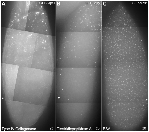
Figure 4. Injected collagenase enzyme disrupts localization to filaments.
Oocytes are positioned with their anterior ends at the top, and injection sites are indicated by asterisks. As the oocyte is too large to be imaged in its entirety at this magnification, images are composites of multiple image stacks, acquired and combined using the Panels and Stitch functions in SoftWoRx. GFP-Mps1 is shown; GFP-Polo responded similarly (data not shown). 4A: Injection of a crude collagenase (Type IV, Gibco) prevented the localization of GFP-Mps1 to filaments in the region around the injection site under hypoxia. 4B: Injection of high purity Clostridiopeptidase A enzyme (Sigma-Aldrich) also prevented the localization of GFP-Mps1 to filaments after exposure to CO2 in the region surrounding the injection site (asterisk). 4C: Control injection of water (data not shown) or a protein solution (5 mg/ml BSA) did not disrupt localization of GFP-Mps1 to the filaments.

