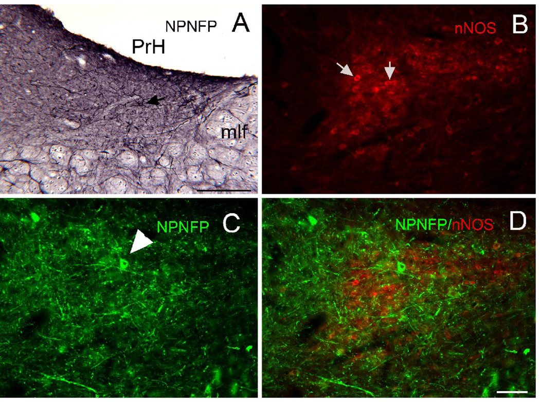
Figure 5.
A. Scattered cells (example at arrow) in PrH are labeled for NPNFP. DAB-GO. Scale bar 250 µm. mlf, medial longitudinal fasciculus. B. Triangular region of nNOS immunoreactivty in PrH. Arrows indicate two labeled cells. C. NPNFP in cells and dendrites in PrH. The arrowhead indicates a labeled cell. D. Merged image showing that the NPNFP cell is found within the nNOS cell cluster but does not colocalize nNOS. Scale bar 100 µm (B, C, D).

