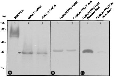
Figure 3.
(A) Profiles of in vitro-translated products. Two different cDNA clones yield identical ≈33-kDa products (lanes 2 and 3). The ≈61-kDa product in lane 1 is generated from the control plasmid. (B) SDS/PAGE of fusion proteins generated from two different constructs. Identical ≈35-kDa bands are seen in gel stained with Coomassie blue. Additional ≈2.5 kDa is caused by the c-myc-(His)6-tag in the fusion protein. (C) Western blot analyses of the fusion protein before (lane 1) and after (lane 2) absorption with the anti-RSOR antibody. The intensity of the ≈35-kDa band is notably reduced after absorption, suggesting that the antibody is specifically directed against the ≈35-kDa RSOR.

