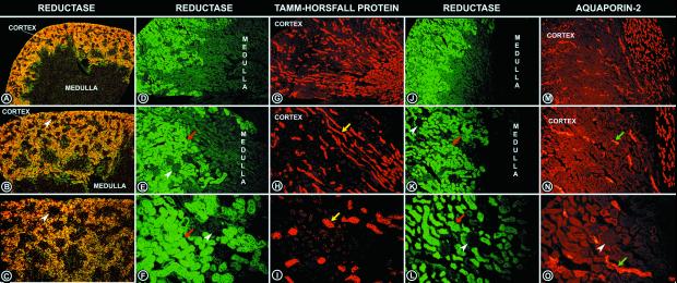
Figure 5.
Low (A), medium (B), and high (C) magnification photomicrographs of in situ autoradiograms of kidney tissue sections hybridized with RSOR riboprobe. The RSOR mRNA is exclusively expressed in tubules of the renal cortex and is absent in the medulla and glomeruli (arrowheads). (D–F and J–L) Photomicrographs with different magnifications of the kidney sections stained with anti-RSOR antibody. The spatial protein expression of RSOR is similar to the mRNA message, and it is absent in the medulla and glomeruli (arrowheads). (G–I) Immunofluorescence photographs of kidney sections stained with anti-Tamm–Horsfall protein antibody, a marker of distal tubular epithelium (arrows). The photographs depicted in G–I are the serial tissue sections of micrographs shown in D–F. (M–O) Immunofluorescence photographs of kidney sections stained with anti-aquaporin-2 antibody, a marker of collecting duct epithelium (arrows). The photographs depicted in M–O are the serial tissue sections of micrographs shown in J–L. Absence of RSOR immunoreactivity in the distal and collecting tubules suggests that it is exclusively expressed in the proximal tubules. Magnifications: A, D, G, J, and M, ×10; B, E, H, K, and N, ×20; C, F, I, L, and O, ×40.

