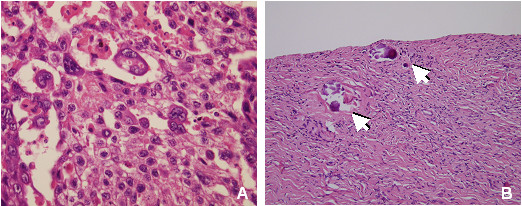
Figure 1.

Histological analysis. Section of choriocarcinoma, left ovary (A): biphasic pattern of sheets of mononuclear cytotrophoblasts and multinuclear syncytiotrophoblasts located near hemorrhagic and necrotic areas. There are no elements suggesting other germ cell tumors (HE staining). Right ovary section (B): fibrous parenchyma, absence of follicles and neoplastic alterations. Arrows: microcalcifications (HE staining).
