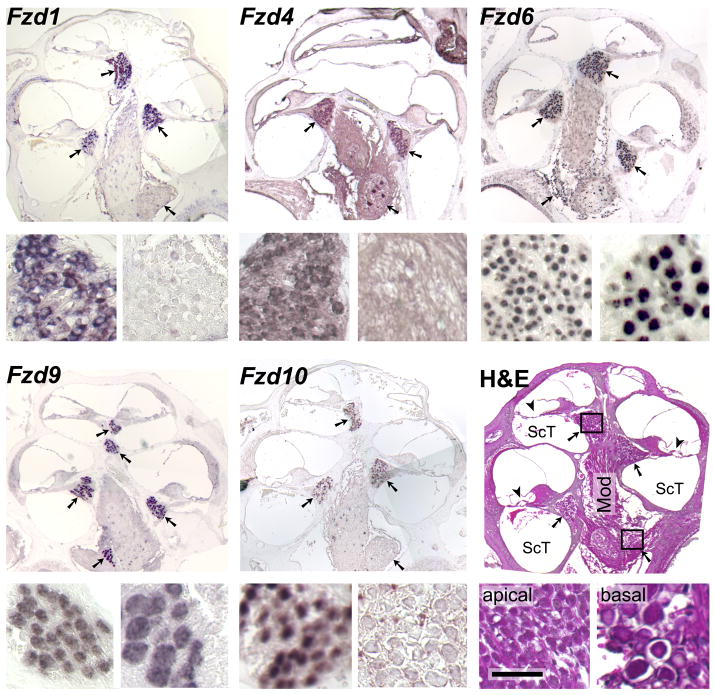
Fig. 3. Selective expression of Wnt receptors in adult spiral ganglion neurons.
In situ hybridizations of antisense riboprobes for the indicated mRNAs to longitudinal paraffin sections of cochleae from two-month old mice, with the apex at the top. Arrows point at the cross-sections of the spiral ganglion inside Rosenthal’s canal; note that some cross sections lack the brown or purple hybridization signal. The smaller panels at the bottom-left and -right of each triad show details from the apical- and basal-most cross sections, respectively, as indicated for the sample stained with hematoxylin and eosin (H&E). Antisense probes for the remaining six Wnt receptors did not exhibit specific labeling, nor did any of the sense probes (data not shown). Mod, modiolus; ScT, scala tympani; arrowheads, location of the organ of Corti with hair cells; scale bar, 300 μm for the large and 50 μm for the small panels.

