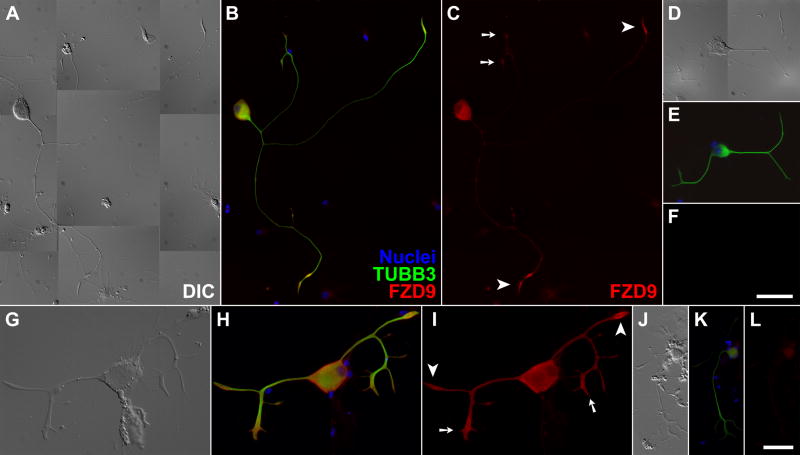
Fig. 6. FZD9 protein localized to growth cones of regenerating adult spiral ganglion neurons in primary culture.
(A/D/G/J) Differential interference contrast (DIC); (B/E/H/K) three-channel epifluorescence with nuclei colored in blue, the neuronal marker βIII-tubulin in green (TUBB3), and FZD9 in red; (C/F/I/L) red-channel fluorescence alone. Only weak background labeling was observed in non-neuronal cells, or when the FZD9 antibodies were omitted (D–F) or replaced by normal rabbit IgG (J–L). Arrowheads, “simple” growth cones; arrows, “complex” growth cones; scalebar in F for panels A-F, 50 μm; scalebar in L for panels G–I and J–L, 17.5 and 30 μm, respectively. The samples in A–F were processed and imaged under identical conditions, as were those in G–L.

