
Figure 4. Intracellular accumulation of the γ2 subunit in organotypic hippocampal slice cultures.
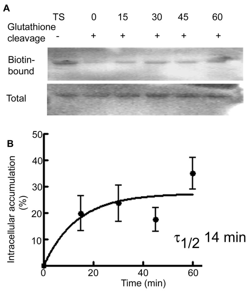
Intracellular accumulation of the γ2 subunit was studied using a biotinylation assay. A representative western blot shows biotin-bound proteins representing the surface expressed fraction of the γ2 subunit (Lane TS) and the amount of receptor accumulated in the intracellular compartment after incubation for 0, 15, 30, 45 and 60 min. Total expression of the γ2 subunit in tissue lysates was similar. B- A ratio of intracellular to surface γ2 subunit was plotted as a function of time (n=4).