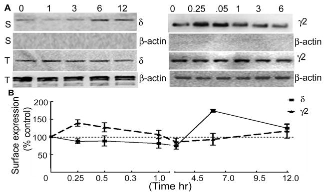
Figure 7. BDNF increased surface expression of the δ subunit of GABARs.
A- Effect of BDNF on surface expression of the δ and γ2 subunits was studied in hippocampal slice cultures. Surface (S) and total (T) expression of the δ and γ2 subunits was studied in slices treated for 1, 3, 6 and 12 hr and 0.25, 0.5, 1, 3 and 6 hr respectively. Surface expression of the δ subunit was higher in slices treated for 6 and 12 hr. On the other hand surface expression of the γ2 subunit was higher at 15 and 30 min. Total expression of the δ and γ2 subunits was similar in all the samples. Blots were re-probed with β-actin antibody and absence of β-actin signal in the surface proteins confirmed specificity of the biotinylation reaction. B- Surface expression of the δ or γ2 subunits in BDNF treated slices was plotted as a percent fraction of surface expressed δ or γ2 subunits in BDNF untreated slices (n=3–4).

