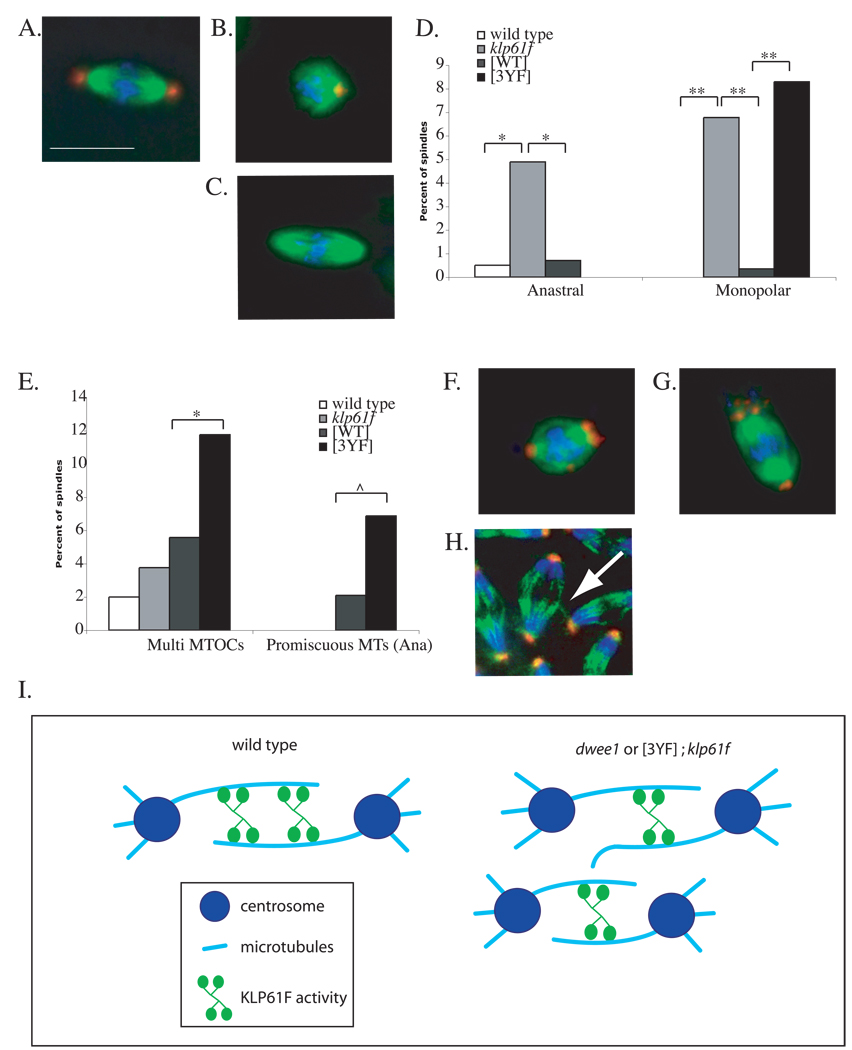
Figure 4. Mitotic spindle defects in embryos containing KLP61FWT or KLP61F3YF.
Embryos from mothers that are wild type, klp61f3 heterozygotes (klp61f), klp61f3 heterozygotes with Myc-KLP61FWT ([WT]) and klp61f3 heterozygotes with Myc-KLP61F3YF ([3YF]) were fixed and stained for DNA (blue), α-tubulin (green), and centrosomin (red). Representative images show a normal spindle (A) and defects significantly different among the genotypes: a monopolar spindle (B), an anastral spindle (C), and spindles with multiple MTOCs (F–G) from interior divisions, and a promiscuous microtubule interaction during anaphase in cortical divisions (H). Scale bar = 5 µm. Defects in (A–C) and (F–H) are quantified in (D) and (E) respectively. See Table 1 for numbers. * p < 0.01, ** p < 0.001, and ^ p < 0.05. (I) A model for regulation of KLP61F to maintain mitotic spindle integrity and prevent promiscuous MT interactions. dWee1 may regulate KLP61F activity to bundle parallel (not shown) and/or anti-parallel microtubules to create a more robust spindle (left). Without dWee1 regulation, KLP61F activity is reduced on the spindle, leading to an unstable spindle with microtubule spurs. These microtubule spurs can then interact with neighboring spindles in a syncytium (spur-spindle interaction is not depicted). Reduced KLP61F activity on the spindle is depicted as reduced protein levels on the spindle, but it remains possible that similar level of protein associate with the spindle but display reduced activity.

