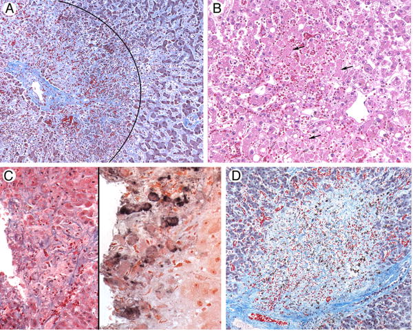
Figure 1.
Photomicrographs of liver tissue illustrating causes of severe hepatitis following hematopoietic cell transplant.
A. Sinusoidal Obstruction Syndrome. A liver acinus is shown here, with a central vein on the left; diffuse hepatocyte necrosis in zone 3, surrounding the vein (the area within the arc); and deposition of collagens within sinusoids in zone 2 of the acinus (outside the arc). Masson trichrome.
B. Hypoxic hepatitis following respiratory failure and hypotension. Zones 2 and 3 of a liver acinus, with the central vein in the lower right. Numerous hepatocytes that have lost nuclei as a result of ischemia are seen in zone 3; arrows point to some of these dying cells. Hematoxylin and eosin.
C. Varicella zoster virus infection. The left panel illustrates a punched-out area of confluent hepatocyte necrosis without distinguishing features (hematoxylin and eosin); the right panel shows reaction product from staining with a monoclonal antibody to VZV (immunohistochemistry).
D. Unknown. There is a large circular area of confluent hepatocyte necrosis surrounded by normal hepatocytes. There were no areas of viral cytopathic effect, and immunohistochemical stains were negative for viruses. Masson trichrome.

