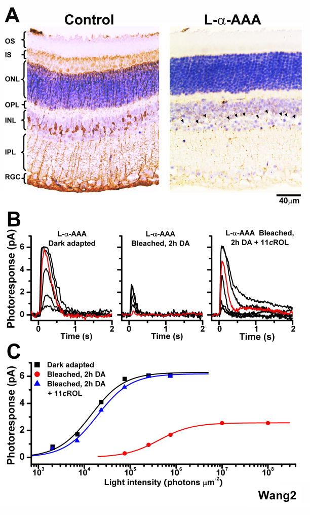
Figure 2.
Effect of the Müller cell inhibitor L-α-AAA on the recovery of mouse cone sensitivity following a bleach
(A) Comparison of the morphology of control retina (left) and retina incubated in 10 mM L-α-AAA gliotoxin for 2.5 h (right). Missing Müller cell nuclei in right panel are indicated by arrowheads
(B) Cone suction recordings from isolated Trα−/− retina pretreated with 10 mM L-α-AAA in darkness for 2.5 h and then transferred to control solution prior to recordings. Cone test-flash responses from retina in dark-adapted state (left), bleached and incubated in darkness for 2 h (middle), and bleached and incubated in darkness for 2 h in the presence of 11-cis retinol (11cROL, right). Red traces represent photoresponses to 75,814 photons μm−2, 500 nm
(C) Intensity-response relations of the cells from (B) fitted with Michaelis-Menten function. The recovery of sensitivity and amplitude of cones from isolated retina were blocked by the gliotoxin, but brought back by the addition of 11-cis retinol.

