
Fig. 1.
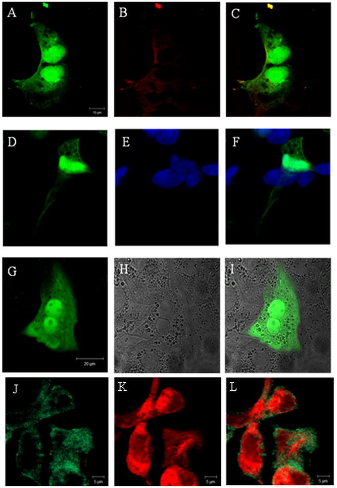
Confocal immunofluorescence shows that (a) PfRACK and (b) RACK1 partially co-localizes in HEK293 cells. (c) Merged image. HEK293 cells (d) expressing GFP-PfRACK and (e) labeled with TO-PRO-3 show that (f) some PfRACK is in the nucleus. Transfection of rat hepatocytes with GFP-PfRACK shows the distribution of this protein is similar in these cells: (g) GFP fluorescence; (h) transmitted light image; (i) merged image. Mz-Cha-1 cells (j) expressing GFP-PfRACK and (k) labeled with an anti- type III InsP3R antibody show that (l) PfRACK only partially co- localizes with this InsP3R isoform.