
Figure 3.
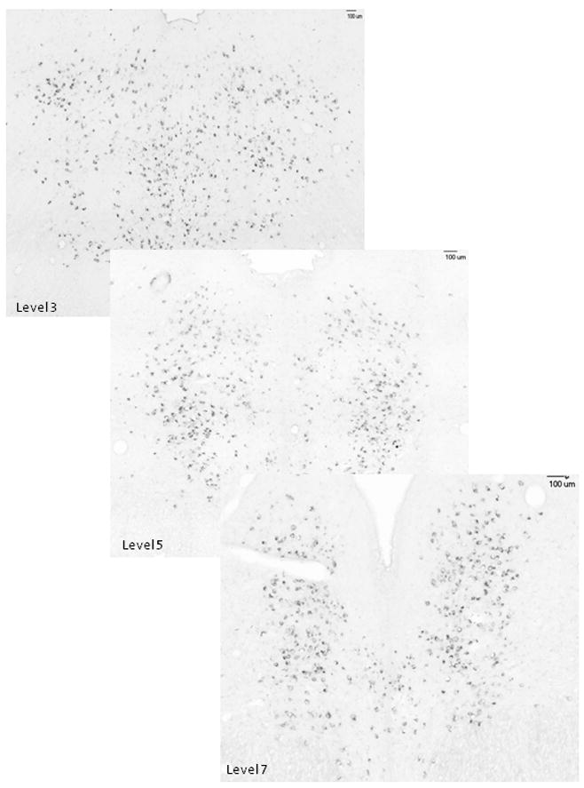
Illustration of the anatomical organization of the Fev-positive neurons at 3 of the 8 levels of the dorsal raphe that were subjected to analysis. The anatomy of the dorsal raphe changes from a unified mass of Fev-positive cells at the rostral levels to a bifurcated structure at the caudal levels.