
Fig. 7.
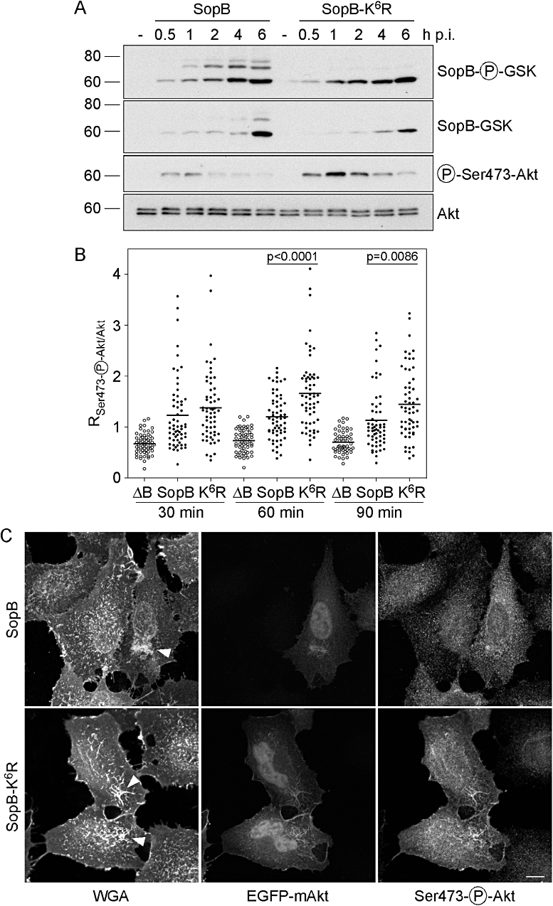
Ubiquitination of SopB downmodulates its activity at the plasma membrane. A. HeLa cells were infected with ΔsopB-sigE Salmonella complemented with either pWSKDE-GSK or pWSKDE-K6R-GSK. At the indicated times, monolayers were solubilized and proteins subject to immunoblotting with antibodies against phospho-GSK (translocated SopB), GSK tag (total SopB), phospho-Ser473-Akt (activated Akt) and Akt pan (total Akt). B. HeLa cells expressing EGFP-mAkt were infected with ΔsopB-sigE Salmonella (ΔB), or ΔsopB-sigE Salmonella complemented with pWSKDE (SopB) or pWSKDE-K6R (K6R). Monolayers were fixed, incubated with Alexa Fluor®647-conjugated WGA to label the plasma membrane, permeabilized and immunostained with anti-phospho-Ser473-Akt antibodies. Single optical sections through individual ruffles were acquired with a spinning disk confocal microscope. Regions of interest were selected by thresholding on the WGA image and transferred to the EGFP-mAkt and phospho-Ser473-Akt images. The ratio of mean fluorescence intensity for phospho-Akt : total Akt was subsequently determined. Each data point represents one ruffle. Data with means from three independent experiments are shown (n ≥ 55 ruffles for each condition). P-values were determined by Student's t-test. C. Representative confocal microscopy images from (B) at 60 min p.i. Arrowheads indicate Salmonella-induced membrane ruffles. Scale bar is 10 μM.