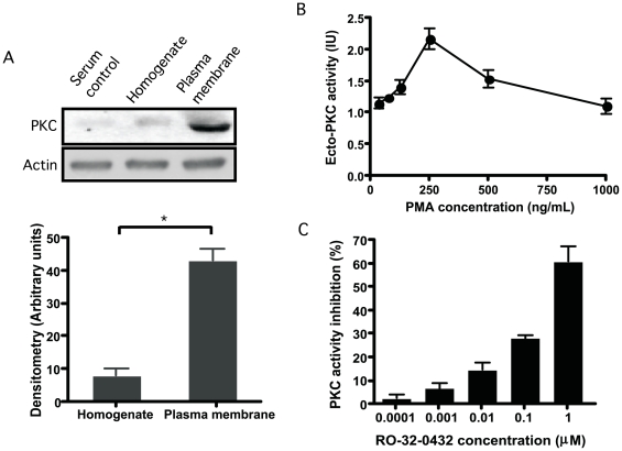
Figure 1. Protein kinase C-like detection on stationary promastigotes.
(A): Immunoblot detection of PKC-like in subcellular fractions of stationary promastigotes of L. mexicana. Cell pellets (2.5×10⁹ cells/ml) were resuspended in 0.02 M Tris-HCl supplemented with 0.25 M sucrose and 3 mM MgCl2. Cells were homogenized and sonicated in a Branson Sonifier 250 W, then resuspended in a buffer containing protease inhibitors and centrifuged at 100000 g for 1 hour at 4°C. Membrane fraction (pellets) and whole homogenates were directly used for immunoblot detection of PKC using a mouse monoclonal anti-PKCα,βI,βII antibody. (B): Ecto-PKC-like activity on intact stationary promastigotes of Leishmania. Promastigotes of L. mexicana were resuspended in 25 mM Tris-HCl, pH 7.0, 3 mM MgCl2, 0.1 mM ATP, 2 mM CaCl2, 50 µg/ml phosphatidylserine, 1 mM EGTA, 0.5 mM EDTA, 0.1 mM PMSF, 1 mM benzamidine. Phorbol myristate acetate (PMA) was added to cell suspensions in concentration ranging from 0.035 to 1 µg/ml and incubated for 15 min. at 30°C. After washes, promastigotes were incubated for 30 min at 30°C with the biotinylated antibody 2B9. PKC activity was revealed by a peroxidase-conjugated streptavidin antibody coupled to ortho-phenyldiamine substrate. Plates were read at 492 nm. Results of three different experiments were expressed in international units (IU/mg) of PKC ± SD. (C): Ecto-PKC-like inhibition. In some experiments intact stationary promastigotes were pretreated with the PKCαβIε inhibitor RO-32-0432 in concentration ranging from 0.0001 to 1 µM during 1 hour at 26°C. PKC activity was revealed by incubating with the biotinylated antibody 2B9. Results of three different experiments were represented as the percentage of PKC activity inhibition ± SD.

