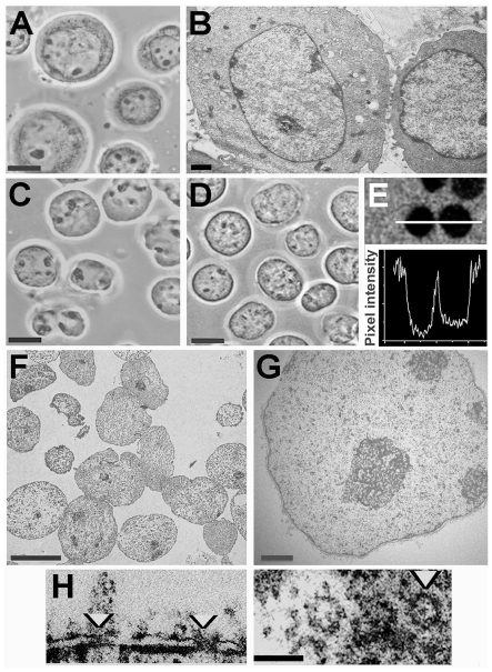
Figure 1. Nuclei of K562 cells released in polymer solution containing 100 µM K-Hepes.
Cells suspended in 70 kDa Ficoll (50% w/v) or 70 kDa dextran (35% w/v) containing digitonin were vortexed to disperse cytoplasmic material, and nuclei were centrifuged and fixed in the same polymer solution. (A) intact cells in growth medium, phase contrast. (B) intact cells, electron microscope section. (C, D) nuclei isolated in Ficoll or dextran respectively, phase contrast. (E) nuclei incubated as in D in dextran with addition of fluorescein-labelled dextran of the same size, fluorescence image of an approximately midplane 0.5 µm confocal section after 1 h; lower panel shows a scan of fluorescence intensity along the white line. (F, G) nuclei isolated in Ficoll and processed as in B, electron microscope images of sections. (H) nuclear pores (arrowheads) in nuclei isolated in Ficoll seen in transverse (left) and tangential (right) electron microscope sections. Bars 10 µm (A, C, D, F); 1 µm (B, G); 0.2 µm (H).

