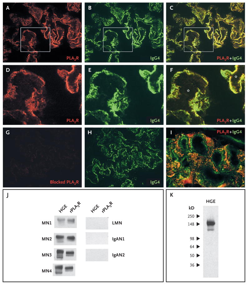
Figure 5. Colocalization of the M-Type Phospholipase A2 Receptor (PLA2R) and IgG4 and Reactivity of Eluted IgG4.
Confocal microscopic analysis of cryosections of a kidney-tissue specimen from a patient with membranous nephropathy revealed the presence of PLA2R (Panel A) and IgG4 (Panel B), which were colocalized in the peripheral capillary walls and the glomerular basement membrane (Panel C). Panels D, E, and F are enlarged images of the boxed areas in Panels A, B, and C, respectively. The asterisks in Panel F indicate the capillary lumina. The anti-PLA2R antibody was blocked with the recombinant fragment (Panel G), with staining virtually eliminated, despite the continued presence of IgG4 (Panel H). Panel I is a confocal image of representative glomerular capillary loops in a biopsy specimen from a patient with lupus membranous nephropathy; PLA2R and IgG4 are not colocalized. IgG4 was detected with sheep anti–human IgG4 antibody (1:500 dilution) and rabbit anti–sheep IgG antibody (1:500 dilution). IgG was eluted from biopsy cores from patients with membranous nephropathy (MN), lupus membranous nephropathy (LMN), or IgA nephropathy (IgAN). This eluted IgG was used to immunoblot human glomerular extract (HGE) or recombinant PLA2R (rPLA2R) (Panel J). Only IgG eluted from the MN samples identified the native and recombinant PLA2R. Panel K shows that the IgG eluted from the MN3 biopsy sample recognized only those bands corresponding to PLA2R.

