Figure 3.
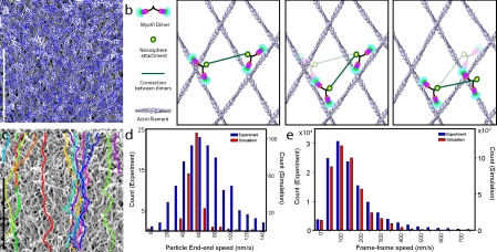
Movement of myosin VI artificial dimer-coated nanospheres on extracted keratocyte actin network assessed from experimentation and simulation. (a) A digitized interlaced actin network used in the simulation (blue) overlaid on the platinum replica micrograph from which it was obtained. (b) A schematic drawing of the simplified model used for the simulation. (b, left) Two dimers bound to actin filaments, coupled through the nanosphere (green line), and bound to the nanosphere at the yellow circle. (b, center) The dimer on the left steps first to its next binding site. (b, right) The dimer on the right steps to the next binding site. Binding sites are selected to maintain the distance between the nanosphere attachment points (length of the green line). (c) Sample trajectories of simulated movement of nanospheres on the extracted keratocyte lamellipodium. (d) Comparison of end–end speeds from the experiment (blue) and simulation (red). (e) Comparison of frame–frame speeds between the experiment (blue) and simulation (red). Bars, 1 µm.

