Figure 1.
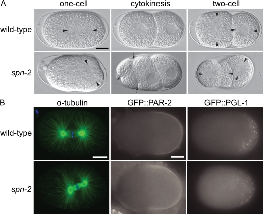
SPN-2 is required for proper spindle positioning. (A) DIC images of live mitotic embryos. Arrowheads mark the centrosomes and arrows point to ectopic cleavage furrows. (B) Confocal micrographs of α-tubulin (green) and DAPI (blue) staining of one-cell metaphase embryos; epifluorescence images of embryos expressing GFP::PAR-2 or GFP::PGL-1. Bars, 10 µm.

