Figure 2.
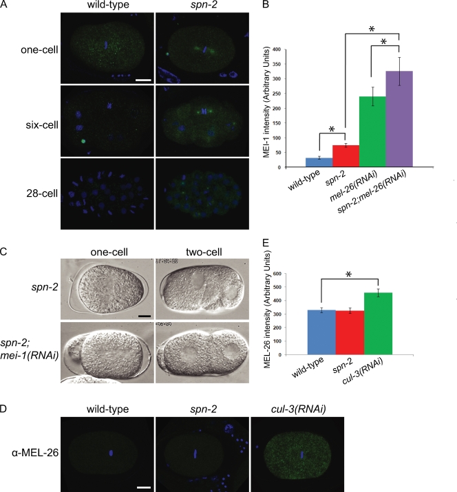
MEI-1 ectopically localizes to mitotic spindles in spn-2 embryos. (A) Confocal micrographs of α-MEI-1 (green) and DAPI (blue) staining. (B) Quantification of spindle MEI-1 levels in one-cell metaphase embryos. (C) DIC images of one-cell anaphase (left) and two-cell interphase (right) embryos. (D) Confocal micrographs of α-MEL-26 (green) and DAPI (blue) staining. (E) Quantification of MEL-26 levels in one-cell embryos. Error bars represent SEM; asterisks indicate statistical significance (P < 0.05). Bar, 10 µm.

