Figure 3.
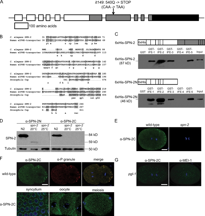
spn-2 encodes an eIF4E-binding protein that localizes to the cytoplasm and P granules. (A) Diagram of spn-2 intron/exon structure and the it149 mutation. White, protein coding sequence; gray, Q-rich domain; black, 3′ UTR. (B) Alignments of the N-terminal region of C. elegans SPN-2 with human 4E-T and an internal region of Drosophila Cup; these regions are 27% and 37% identical, respectively, to SPN-2 (identities shaded). The eIF4E-binding motif (YxxxxLϕ, where ϕ is a hydrophobic residue) is boxed. (C) 6xHis-tagged SPN-2 or SPN-2N were incubated with GST-eIF4E isoforms; eluates were analyzed by Western blotting with α-6xHis. (D) Western blots of wild-type (N2) and spn-2 worm extracts probed with α-SPN-2N or α-SPN-2C and reprobed with α-tubulin as a loading control. (E–G) Confocal images of wild type or mutants stained with the indicated antibodies and DAPI (blue). (E) One-cell embryos. (F, top) Two-cell embryos. (F, bottom) Wild-type germ-line syncytium, oocytes, and a meiotic embryo. (G) One-cell embryos. Bars, 10 µm.

