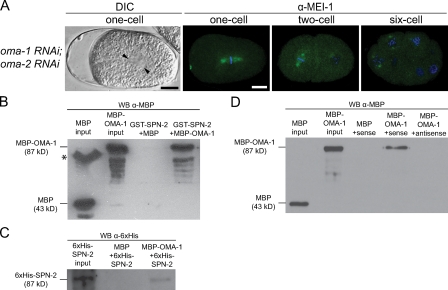
Figure 4.
OMA-1 interacts with SPN-2 and the mei-1 3′ UTR. (A) DIC image of a metaphase embryo with skewed spindle (arrowheads mark the centrosomes), and confocal images of embryos stained for MEI-1 (green) and DAPI (blue). Bar, 10 µm. (B and C) In vitro pull-down assays using the proteins shown; eluates were analyzed by Western blotting (WB) with the indicated antibodies. The asterisk in B indicates a band of aggregated MBP in the MBP input lane. (D) MBP–OMA-1 or MBP was incubated with biotinylated sense or antisense RNA from the mei-1 3′ UTR. RNA-bound proteins were analyzed by Western blotting.

