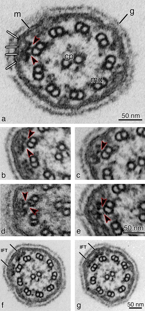
Figure 4.
TEM images of cross sections of flagella from flat-embedded pf28 mutant C. reinhardtii cells, which lack outer dynein arms. (a) Cross section of pf28 flagella showing IFT trains located underneath the flagellar membrane (m). The arrowheads indicate the two links connecting the bilobed IFT particle to the B tubule of a MT doublet (md). The same structures can be observed in b–e (arrowheads). The IFT particle also shows links to the flagellar membrane (white lines). cp, central pair; g, glycocalyx (a membrane overlay composed of hydroxyproline-rich glycoproteins, which surrounds the C. reinhardtii flagella). (b–e) Cross sections showing bilobed IFT particles. Each IFT particle has connections with the flagellar membrane on one side and connections to the B tubule of the MT doublet on the other side (arrowheads). (f and g) Two consecutive serial cross sections of a flagellum from a pf28 mutant cell showing two side-by-side IFT trains on adjacent doublet MTs (arrows). (g) Adjacent IFT trains can occasionally contact each other laterally, forming wider structures that can extend for ∼70 nm over two neighboring MT doublets.

