Figure 3.
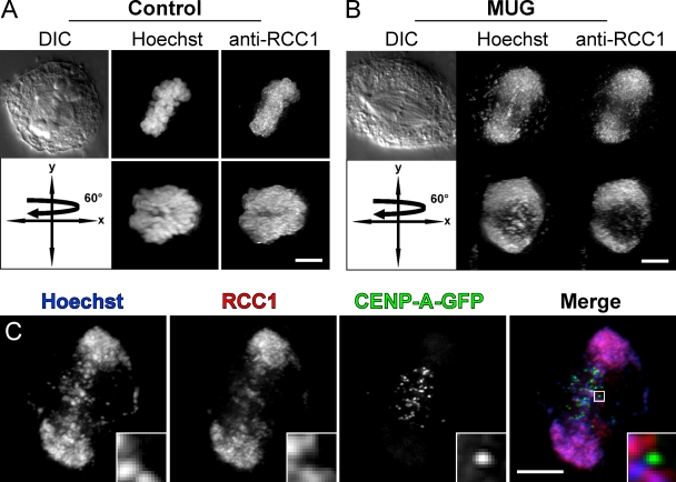
RCC1 binds to chromatin but not kinetochores during MUG. (A) In a normal mitotic cell, the RanGEF RCC1 is evenly distributed on chromosomes. A view rotated by 60° around the y axis shows a uniform coating through the entire volume at which the mitotic spindle assembles. (B) During MUG, RCC1 is also bound to chromatin but is pushed to the side by the spindle that assembles around kinetochores. This is most evident as a hole created by spindle microtubules passing through the chromatin visible in the rotated view. (C) Although antibodies against RCC1 recognize small pieces of equatorial chromatin, centromeres/kinetochores labeled with CENP-A–GFP lack RCC1 staining (insets). Bars, 5 µm.

