Figure 6.
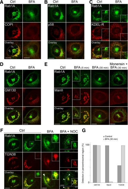
The mobility of the pcIC facilitates protein localization. Control NRK cells expressing GFP-Rab1A, and cells treated for different times (5 or 30 min) with BFA, were stained with antibodies against β-COP (A), p58 (B), KDEL-R (C), GM130 (D), mannosidase II (E), and TGN38 (F). In addition, the right panels in E and F show the localizations of mannosidase II on TGN38 in cells treated for 30 min with a combination of BFA + monensin or BFA + nocodazole, respectively. (G) Determination of the percentage of cells showing pericentrosomal labeling for GM130, mannosidase II and TGN38 (n = 250). Note that the cells were simply scored as positive or negative, irrespective of the intensity of the fluorescent signals, which were highly variable. Bars, 10 μm.

