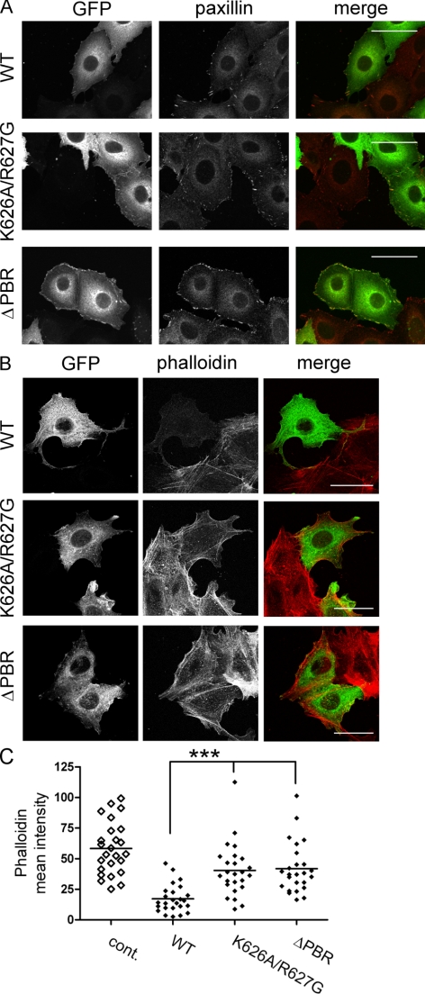
Figure 6.
The PBR is required for DLC1-mediated actin cytoskeletal changes. (A and B) MCF7 cells grown on collagen-coated coverslips were transfected with expression plasmids encoding GFP-DLC1 WT, K626A/R627G, and ΔPBR and fixed after 24 h. (A) Focal adhesions were visualized with a paxillin-specific primary and Alexa Fluor 546–coupled secondary antibody (red). (B) Actin stress fibers were stained with Alexa Fluor 546–conjugated phalloidin (red). The images shown are stacks of five to six confocal sections taken at 0.5-μm intervals. Scale bars, 50 μm. (C) The mean phalloidin intensity of cells expressing similar levels of the different GFP-DLC1 variants (n > 25) was quantified. Nontransfected cells were used as a control (cont.).

